Skin lesions are areas of the skin that look different from the surrounding skin. They can range from harmless moles to more serious growths that may require medical attention. Knowing the difference between benign (non-cancerous) and malignant (cancerous) lesions is important for early detection and effective treatment.
Benign lesions usually grow slowly, stay clearly defined, and do not spread to other areas. Examples include seborrhoeic keratoses, dermatofibromas, and common moles. These are generally not dangerous unless they become irritated or affect appearance.
Malignant lesions are cancerous and can grow quickly, invade surrounding tissue, and spread to other parts of the body. Common types include basal cell carcinoma, squamous cell carcinoma, and melanoma. Early detection is crucial for better outcomes.
.jpg)
The ABCDE rule is a helpful guide to spot potential warning signs, especially for melanoma:
Even if a lesion does not fit all these criteria, any change should be assessed by a healthcare professional.
Some harmless growths can resemble cancer, which is why professional assessment is important:
Seek prompt evaluation if a lesion shows:
Early consultation allows for accurate diagnosis and treatment, reducing the risk of complications.
Treatment depends on the type of lesion and its location:
Benign lesions:
Malignant or suspicious lesions:
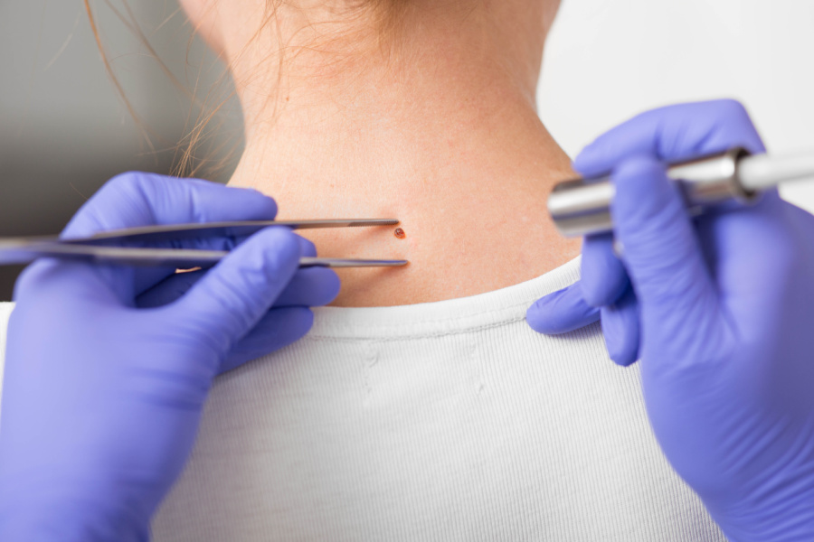
Minor skin surgery is often safe, quick, and effective for removing problematic lesions.
Routine self-checks and professional skin assessments help catch issues early.
Understanding your skin and spotting changes early can make a real difference. If you notice a suspicious lesion or one that is changing, it is best to get it checked promptly. The Minor skin surgery clinic offer safe and effective options for removing benign or concerning lesions with minimal downtime.
Book a consultation today to have your skin assessed and ensure your skin health is protected.